Ancient ozone holes may have sterilized forests 252 million years ago

Volcano-fueled holes in Earth’s ozone layer 252 million years ago may have repeatedly sterilized large swaths of forest, setting the stage for the world’s largest mass extinction event. Such holes would have allowed ultraviolet-B radiation to blast the planet. Even radiation levels below those predicted for the end of the Permian period damage trees’ abilities to make seeds, researchers report February 7 in Science Advances.

Jeffrey Benca, a paleobotanist at the University of California, Berkeley, and his colleagues exposed plantings of modern dwarf pine tree (Pinus mugo) to varying levels of UV-B radiation. Those levels ranged from none to up to 93 kilojoules per square meter per day. According to previous simulations, UV-B radiation at the end of the Permian may have increased from a background level of 10 kilojoules (just above current ambient levels) to as much as 100 kilojoules, due to large concentrations of ozone-damaging halogens spewed from volcanoes (SN: 1/15/11, p. 12).

Exposure to higher UV-B levels led to more malformed pollen, the researchers found, with up to 13 percent of the pollen grains deformed under the highest conditions. And although the trees survived the heightened irradiation, the trees’ ovulate cones — cones that, when fertilized by pollen, become seeds — did not. But the trees weren’t permanently sterilized: Once removed from extra UV-B exposure, the trees could reproduce again.

The finding supports previous research suggesting that colossal volcanic eruptions in what’s now Siberia, about 300,000 years before the onset of the extinction event, probably triggered the die-off of nearly all marine species and two-thirds of species living on land (SN: 9/19/15, p. 10). Repeated pulses of volcanism at the end of the Permian may have led to several periods of irradiation that sterilized the forests, causing a catastrophic breakdown of food webs, the researchers say — an indirect but effective way to kill.

This stick-on patch could keep tabs on stroke patients at home

AUSTIN, Texas — Stretchy sensors that stick to the throat could track the long-term recovery of stroke survivors.

These new Band-Aid‒shaped devices contain motion sensors that detect muscle movement and vocal cord vibrations. That sensor data could help doctors diagnose and monitor the effectiveness of certain treatments for post-stroke conditions like difficulty swallowing or talking, researchers reported February 17 in a news conference at the annual meeting of the American Association for the Advancement of Science. Up to 65 percent of stroke survivors have trouble swallowing, and about a third of survivors have trouble carrying on conversations.
The devices can monitor speech patterns more reliably than microphones by sensing tissue movement rather than recording sound. “You don’t pick up anything in terms of ambient noise,” says study coauthor John Rogers, a materials scientist and bioengineer at Northwestern University in Evanston, Ill. “You can be next to an airplane jet engine. You’re not going to see that in the [sensor] signal.”

Developed by Rogers’ team, the sensors have built-in 12-hour rechargeable batteries and continually stream motion data to a smartphone. Researchers are now testing the sensors with real stroke patients to see how the devices can be made more user-friendly. For instance, Rogers’ team realized that patients were unlikely to wear sensors that were too easily visible. By equipping the patches with more sensitive motion sensors, they can be worn lower on a person’s neck, hidden behind a buttoned-up shirt, and still pick up throat motion.

These kinds of sensors could also track the recovery of neck cancer patients, who commonly develop swallowing and speaking problems caused by radiation therapy and surgery, Rogers says. The devices can also measure breathing and heart rates to monitor sleep quality and help diagnose sleep apnea. Rogers expects this wearable tech to be ready for widespread use within the next year or two.

Some flu strains can make mice forgetful

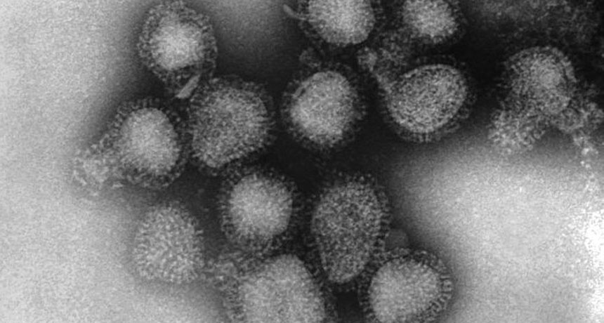
With fevers, chills and aches, the flu can pound the body. Some influenza viruses may hammer the brain, too. Months after being infected with influenza, mice had signs of brain damage and memory trouble, researchers report online February 26 in the Journal of Neuroscience.

It’s unclear if people’s memories are affected in the same way as those of mice. But the new research adds to evidence suggesting that some body-wracking infections could also harm the human brain, says epidemiologist and neurologist Mitchell Elkind of Columbia University, who was not involved in the study.
Obvious to anyone who has been waylaid by the flu, brainpower can suffer at the infection’s peak. But not much is known about any potential lingering effects on thinking or memory. “It hasn’t occurred to people that it might be something to test,” says neurobiologist Martin Korte of Technische Universität Braunschweig in Germany.

The new study examined the effects of three types of influenza A — H1N1, the strain behind 2009’s swine flu outbreak; H7N7, a dangerous strain that only rarely infects people; and H3N2, the strain behind much of the 2017–2018 flu season misery (SN: 2/17/18, p. 12). Korte and colleagues shot these viruses into mice’s noses, and then looked for memory problems 30, 60 and 120 days later.

A month after infection, the mice all appeared to have recovered and gained back weight. But those that had received H3N2 and H7N7 had trouble remembering the location of a hidden platform in a pool of water, the researchers found. Mice that received no influenza or the milder H1N1 virus performed normally at the task.
Researchers also studied the brain tissue of the infected mice under a microscope and found that the memory problems tracked with changes in nerve cells. A month after H7N7 or H3N2 infection, mice had fewer nerve cell connectors called dendritic spines on cells in the hippocampus, a brain region involved in memory. Electrical experiments on the nerve cell samples in dishes also suggested the cells’ signal-sending abilities were impaired.
What’s more, these mice’s brains looked inflamed under the microscope, full of immune cells called microglia that were still revved up 30 and 60 days after infection. Cell counts revealed that mice that had suffered through H3N2 or H7N7 had more active microglia than mice infected with H1N1 or no virus at all. That lingering activity was surprising, Korte says; most immune cells in the body usually settle down soon after an infection clears.

These memory problems and signs of brain trouble were gone by 120 days, which translates to about a decade in human time, Korte says. “I’m not saying that everyone who has influenza is cognitively impaired for 10 years,” he says, noting that human brains are much more complex than those of mice. “The news is more that we should not only look at lung functionality after the flu, but also cognitive effects, weeks and months after infection.”

H7N7 can infect brain cells directly. But H1N1 and H3N2 don’t typically get into the brain (and Korte and colleagues confirmed that in their experiments). Some flu viruses may be causing brain trouble remotely, perhaps through inflammatory signals in the blood making their way into the brain, the study suggests. If that pathway is confirmed, then many types of infections could cause similar effects on the brain. “It is plausible that this is a general phenomenon,” Elkind says.