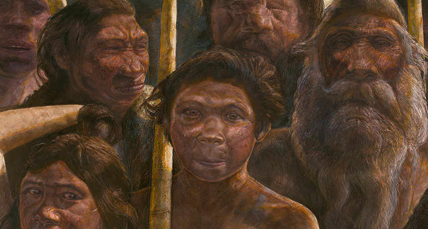
Neandertals hung out in what’s now northern Spain around 430,000 years ago, an analysis of ancient DNA suggests. That’s an earlier Neandertal presence in Europe, by at least 30,000 years, than many researchers had assumed.
Fragments of nuclear DNA from a tooth and partial leg bone discovered at Sima de los Huesos, a chamber deep inside a Spanish cave, resemble corresponding parts of a previously reassembled Neandertal genome, researchers say in a study published online March 14 in Nature. Not much nuclear DNA survives in such ancient fossils, say paleogeneticist Matthias Meyer of the Max Planck Institute for Evolutionary Anthropology in Leipzig, Germany, and his colleagues. Meyer’s group recovered DNA fragments covering a fraction of 1 percent of the newly recovered Neandertal tooth and leg genomes. Just enough DNA remained to enable comparisons with DNA of a Neandertal woman (SN: 1/25/14, p. 17) and a Denisovan woman (SN: 9/22/12, p. 5). Denisovans are considered close genetic cousins of Neandertals.
The early age for the new genetic finds challenges the idea that fossils from Sima de los Huesos, or pit of bones, come from a species called Homo heidelbergensis. Some researchers have suspected that by around 400,000 years ago, H. heidelbergensis gave rise to evolutionary precursors of both Neandertals and Homo sapiens. An ancient genetic puzzle has also emerged at Sima de los Huesos. On one hand, nuclear DNA — which passes from both parents to their children — pegs the Spanish hominids as Neandertals. But mitochondrial DNA — typically inherited only from the mother — already extracted from one Sima de los Huesos fossil (SN: 12/28/13, p. 8) and described for a second fossil in the new study has more in common with Denisovans.
Denisovans lived in East Asia at least 44,000 years ago, but their evolutionary history is unknown.
If early Neandertals lived in northern Spain roughly 430,000 years ago, “we have to go back further in time to reach the common ancestor of Neandertals and Denisovans,” Meyer says. The new genetic data from Sima de los Huesos now suggest that Denisovans split from Neandertals perhaps 450,000 years ago, says paleoanthropologist Chris Stringer of the Natural History Museum in London. Genetic and fossil evidence point to Neandertals and H. sapiens diverging from a common ancestor around 650,000 years ago, he proposes.
But it’s hard to say whether that common ancestor was H. heidelbergensis, Stringer adds. “Research must refocus on fossils from 400,000 to 800,000 years ago to determine which ones might lie on ancestral lineages of Neandertals, Denisovans and modern humans.”
Hominids throughout Eurasia during that time may have shared a mitochondrial DNA pattern observed in Sima de los Huesos Neandertals and Asian Denisovans, Meyer suggests. If that was the case, Neandertals acquired a new form of mitochondrial DNA by interbreeding with modern humans or their direct ancestors from Africa sometime between 430,000 and 100,000 years ago (SN: 3/19/16, p. 6).
Another possibility is that Neandertals traveled to Europe from Asia more than 430,000 years ago, carrying Denisovan mitochondrial DNA with them, says paleogeneticist Carles Lalueza-Fox of the Institute of Evolutionary Biology in Barcelona. Or hybrid descendants of early Neandertals and early Denisovans may have lived at Sima de los Huesos, carrying Denisovan mitochondrial DNA, he speculates.
“We really need more genetic data from Sima de los Huesos, and other sites of that age, to narrow down these scenarios,” Meyer says.
In the 1967 animated Disney film The Jungle Book, the feral boy Mowgli encounters a jazz-singing orangutan named King Louie, who implores Mowgli to teach him the secret of fire. King Louie presented a challenge for the producers of Disney’s live-action, CGI-enhanced remake of the film, opening April 15. “We had this notion that we would be as authentic as we could be to the region,” says producer Brigham Taylor. The problem: Orangutans are not native to India. In fact, King Louie himself is not native to Rudyard Kipling’s original stories. But instead of scrapping the character, the filmmakers got creative. While researching India’s wildlife, the film’s art department learned that a colossal ape named Gigantopithecus once roamed the region. Various species of Gigantopithecus lived in India, China and Southeast Asia from about 6.5 million years ago until as recently as a few hundred thousand years ago. The ape was truly gigantic — by some estimates, twice as big as a gorilla.
So King Louie morphed from orangutan to Gigantopithecus. The switch was a “fun justification,” Taylor says, to keep the character and play up his size while still staying true to India’s fauna. (Yes, the ape is extinct, but this is a movie about talking animals. And fossil evidence does suggest that the ape at least mingled with the human ancestor Homo erectus.)
Using the scientific information they could find on the Internet, visual effects artists imagined how the animal would look and move, Taylor says. The result: an ape that resembles an overgrown orangutan, Gigantopithecus’ closest living relative. The movie ape has shaggy hair, flaring cheeks and a saggy pouch that hangs from the throat like a double chin — and towers about 12 feet tall. It’s difficult to judge how accurate Disney’s rendering is. Despite possibly having been the largest primate ever to have lived, Gigantopithecus left behind few fossils. Scientists have just four lower jaws and over a thousand teeth, says biological anthropologist Russell Ciochon of the University of Iowa. That’s not much to go on, but Ciochon and colleagues made their own reconstruction a couple decades ago. The researchers took a jaw from China and made an outline of a skull that could fit such a jaw. Because most primate skulls scale to body size, Ciochon says, his group could estimate Gigantopithecus’ weight, 800 to 900 pounds, and height, about 9 feet from head to toe. (The species that lived in India was actually probably smaller.) Adding other details like hair to the animal is a matter of conjecture, Ciochon says.
But the teeth do offer some solid details about the ape’s lifestyle. Wear patterns and microscopic debris stuck to the teeth indicate Gigantopithecus dined on fruits, leaves, shoots, roots and perhaps even bamboo. Last year, researchers confirmed those details after analyzing the ratios of carbon isotopes in teeth found in Southeast Asia. The analysis also determined that Gigantopithecus was a strict forest dweller, even though it also lived near grasslands in some areas. In fact, the researchers contend, Gigantopithecus’ reliance on forests and its big size — and therefore big appetite — may have been the animal’s undoing. As Southeast Asia’s jungles gave way to expanding grasslands during the last glacial period, Gigantopithecus may have been unable to cope.
Perhaps if our ancestors had shared the secret of fire with Gigantopithecus, the giant ape would still be around today.
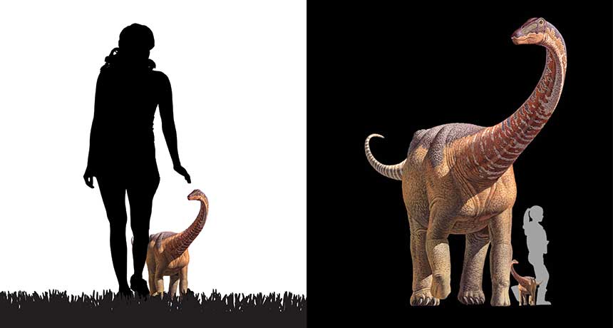
A baby titanosaur looked a lot like a grown-up — and it probably acted like one, too.
The (relatively) tiny fossils of a roughly 1- to 2-month-old dinosaur, Rapetosaurus krausei, discovered in what is now Madagascar, suggest that babies and adults had similar limb proportions, researchers report in the April 22 Science. That’s a sign that the babies were precocious, or didn’t require a whole lot of parental care, says study coauthor Kristi Curry Rogers, a vertebrate paleontologist at Macalester College in St. Paul, Minn. After hatching, she says, the tiny titanosaur may have been more self-reliant than babies of other dinosaur species.
A lack of very young titanosaur specimens has made it tough to understand the enormous dinosaurs’ growth patterns. Curry Rogers and colleagues estimate that when newly hatched, the baby weighed 3.4 kilograms — about the weight of a newborn human. But in just a few weeks, the dinosaur’s weight shot up to 40 kilograms, roughly as heavy as a 12-year-old boy.
During the growth spurt, all of the baby’s limbs grew at about the same rate, the team calculated with data from microscopic images and CT scans. Those data plus features of the bones’ tissue point toward a life that, though cut short by starvation, was both active and independent.
In the summer of 2013, an epidemic began sweeping through the intertidal zone off the west coast of North America. The victims were several species of sea star, including Pisaster ochraceus, a species that comes in orange and purple variants. (It’s also notable because it’s the starfish that provided ecology with the fundamental concept of a keystone species.) Affected individuals appeared to “melt,” losing grip with the rocks to which they were attached — and then losing their arms. This sea star wasting disease, as it is known, soon killed sea stars from Baja California to Alaska.
This wasn’t the first outbreak of sea star wasting disease. A 1978 outbreak in the Gulf of California, for instance, killed so many Heliaster kubinjiisun stars that the once ubiquitous species is now incredibly rare.
These past incidents, though, happened fast and within smaller regions, so scientists had struggled to figure out what had happened. With the latest outbreak happening over such a large — and well-studied — region and period of time, marine biologists have been able to gather more data on the disease than ever before. And they’re getting closer to figuring out just what happened in this latest incident.
One likely factor is the sea star-associated densovirus, which, in 2014, scientists reported finding in greater abundance in starfish with sea star wasting disease than in healthy sea stars. But the virus can’t be the only cause of the disease; it’s found in both healthy and sick sea stars, and it has been around since at least 1942, the earliest year it has been found in museum specimens. So there must be some other factor at play. Earlier this year, scientists studying the outbreak in Washington state reported in the Proceedings of the Royal Society B thatwarm waters may increase disease progression and rates of death. Studies of California starfish came to a similar conclusion. But a new study, appearing May 4 in PLOS One , finds that may not be true for sea stars in Oregon. Bruce Menge and colleagues at Oregon State University took advantage of their long-term study of Oregon starfish to evaluate what happened to sea stars during the recent epidemic and found that wasting disease increased with cooler , not warmer, temperatures. “Given conflicting results on the role of temperature as a trigger of [sea star wasting disease], it seems most likely that multiple factors interacted in complex ways to cause the outbreak,” they conclude. What those factors are, though, is still a mystery.
Also unclear is what long-term effects this outbreak will have on Pacific intertidal communities.
In the 1960s, Robert Paine of the University of Washington performed what is now considered a classic experiment. For years, he removed starfish from one area of rock in Makah Bay at the northwestern tip of Washington and left another bit of rock alone as a control. Without the starfish to prey on them, mussels were able to take over. The sea stars, Paine concluded, were a “keystone species” that kept the local food web in control.
If sea star wasting disease has similar effects on the Pacific intertidal food web, Menge and his colleagues write, “it would result in losses or large reductions of many species of macrophytes, anemones, limpets, chitons, sea urchins and other organisms from the low intertidal zone.”
What happens, the group says, may depend on how quickly the disease disappears from the region and how many young sea stars can grow up and start munching on mussels.
It’s hard to believe that it took reality television this long to get around to dealing with space, time and our place in the cosmos.
In PBS’ Genius by Stephen Hawking, the physicist sets out to prove that anyone can tackle humankind’s big questions for themselves. Each of the series’ six installments focuses on a different problem, such as the possibility of time travel or the likelihood that there is life elsewhere in the universe. With Hawking as a guide, three ordinary folks must solve a series of puzzles that guide them toward enlightenment about that episode’s theme. Rather than line up scientists to talk at viewers, the show invites us to follow each episode’s trio on a journey of discovery. By putting the focus on nonexperts, Genius emphasizes that science is not a tome of facts handed down from above but a process driven by curiosity. After working through a demonstration of how time slows down near a black hole, one participant reflects: “It’s amazing to see it play out like this.” The show is a fun approach to big ideas in science and philosophy, and the enthusiasm of the guests is infectious. Without knowing what was edited out, though, it’s difficult to say whether the show proves Hawking’s belief that anyone can tackle these heady questions. Each situation is carefully designed to lead the participants to specific conclusions, and there seems to be some off-camera prompting.
But the bigger message is a noble one: A simple and often surprising chain of reasoning can lead to powerful insights about the universe, and reading about the cosmos pales next to interacting with stand-ins for its grandeur. It’s one thing, for example, to hear that there are roughly 300 billion stars in the Milky Way. But to stand next to a mountain of sand where each grain represents one of those stars is quite another. “I never would have got it until I saw it,” says one of the guests, gesturing to the galaxy of sand grains. “This I get.”
In hunting down delicious fish, Flipper may have a secret weapon: snot.
Dolphins emit a series of quick, high-frequency sounds — probably by forcing air over tissues in the nasal passage — to find and track potential prey. “It’s kind of like making a raspberry,” says Aaron Thode of the Scripps Institution of Oceanography in San Diego. Thode and colleagues tweaked a human speech modeling technique to reproduce dolphin sounds and discern the intricacies of their unique style of sound production. He presented the results on May 24 in Salt Lake City at the annual meeting of the Acoustical Society of America.
Dolphin chirps have two parts: a thump and a ring. Their model worked on the assumption that lumps of tissue bumping together produce the thump, and those tissues pulling apart produce the ring. But to match the high frequencies of live bottlenose dolphins, the researchers had to make the surfaces of those tissues sticky. That suggests that mucus lining the nasal passage tissue is crucial to dolphin sonar.
The vocal model also successfully mimicked whistling noises used to communicate with other dolphins and faulty clicks that probably result from inadequate snot. Such techniques could be adapted to study sound production or echolocation in sperm whales and other dolphin relatives. Researchers modified a human speech model developed in the 1970s to study dolphin echolocation. The animation above mimics the vibration of lumps of tissue (green) in the dolphin’s nasal passage (black) that are drenched in mucus. Snot-covered tissues (blue) stick together (red) and pull apart to create the click sound.
Jupiter’s turbulence is not just skin deep. The giant planet’s visible storms and blemishes have roots far below the clouds, researchers report in the June 3 Science. The new observations offer a preview of what NASA’s Juno spacecraft will see when it sidles up to Jupiter later this year.
A chain of rising plumes, each reaching nearly 100 kilometers into Jupiter, dredges up ammonia to form ice clouds. Between the plumes, dry air sinks back into the Jovian depths. And the famous Great Red Spot, a storm more than twice as wide as Earth that has churned for several hundred years, extends at least dozens of kilometers below the clouds as well.
Jupiter’s dynamic atmosphere provides a possible window into how the planet works inside. “One of the big questions is what is driving that change,” says Leigh Fletcher, a planetary scientist at the University of Leicester in England. “Why does it change so rapidly, and what are the environmental and climate-related factors that result from those changes?”
To address some of those questions, Imke de Pater, a planetary scientist at the University of California, Berkeley, and colleagues observed Jupiter with the Very Large Array radio observatory in New Mexico. Jupiter emits radio waves generated by heat left over from its formation about 4.6 billion years ago. Ammonia gas within Jupiter’s atmosphere intercepts certain radio frequencies. By mapping how and where those frequencies are absorbed, the researchers created a three-dimensional map of the ammonia that lurks beneath Jupiter’s clouds. Those plumes and downdrafts appear to be powered by a narrow wave of gas that wraps around much of the planet.
The depths of Jupiter’s atmospheric choppiness isn’t too surprising, says Scott Bolton, a planetary scientist at the Southwest Research Institute in San Antonio. “Almost everyone I know would have guessed that,” he says. But the observations do provide a teaser for what to expect from the Juno mission, led by Bolton. The spacecraft arrives at Jupiter on July 4 to begin a 20-month investigation of what’s going on beneath Jupiter’s clouds using tools similar to those used in this study.
The new observations confirm that Juno should work as planned, Bolton says.
By getting close to the planet — just 5,000 kilometers from the cloud tops — Juno will break through the fog of radio waves from Jupiter’s radiation belts that obscures observations made from Earth and limits what telescopes like the Very Large Array can see. But the spacecraft will see only a narrow swath of Jupiter’s bulk at a time. “That’s where ground-based work like the research de Pater has been doing is really essential,” Fletcher says. Observations such as these will let Juno scientists know what’s going on throughout the atmosphere so they can better understand what Jupiter is telling them.
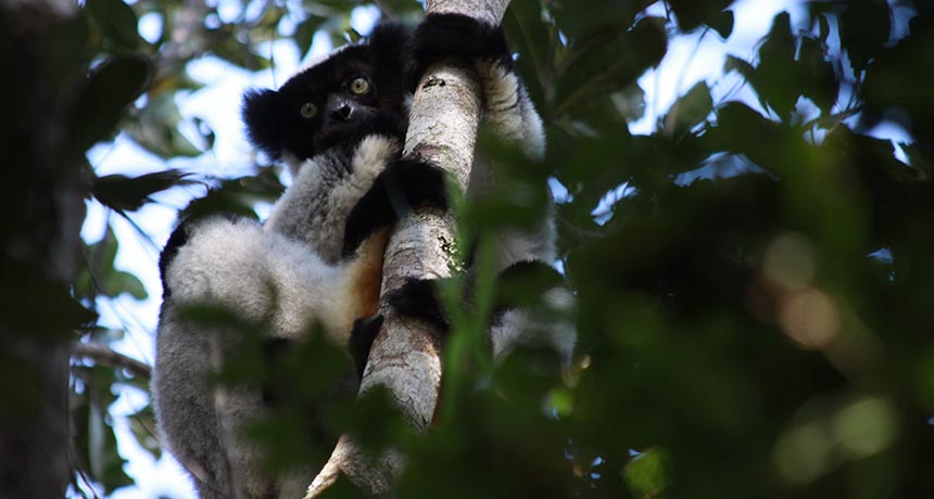
In a chorus of indris, young males vie for the spotlight, riffing in alternation rather than singing in unison. Not content to be the Joey Fatone of the group, these guys strive for Justin Timberlake status.
Indris (Indri indri), the only singing lemur species, begin their songs with roars that descend into long, phrased howls. These choirs are composed of males and females, with one dominant pair. Marco Gamba of the University of Turin in Italy and his colleagues wanted to analyze variation among individual singers.
Listening to 496 indri songs recorded over 10 years in the dense forests of Madagascar, the team found that pitch varies between males and females. And indri groups typically sing in synchrony, amplifying their tunes and vocally marking their territory to other groups. When one singer starts to croon, the others join in and match rhythm.
Solos are rare, but young male singers tend to sing out of sync — probably to stand out and advertise their masculinity, Gamba and his colleagues propose June 14 in Frontiers in Neuroscience.
There are degrees of slothfulness, it turns out, even when it comes to sloths. And three-toed sloths may be the most slothful of them all: A species of the animal has a field metabolic rate that is the lowest ever recorded for any mammal in the world.
Jonathan Pauli, an ecologist at the University of Wisconsin-Madison, got interested in sloths not because they’re adorable but because “other things eat them,” he says. And he stayed interested in the animals because they are “biologically fascinating.”
Sloths are a type of arboreal folivore, a group that includes all animals that live in trees and eat only leaves. What most people lump into the category of “sloth” are really six species in two families (two-toed and three-toed) separated by millions of years of evolution. Both families live in trees in Central and South America and eat leaves, but three-toed sloths tend to have smaller ranges and more constricted diets, eating from only a few species of trees and only a limited number of them. Studies have also shown that these sloths have a very slow metabolic rate.
But how slow? To find out, Pauli and his colleagues captured 10 brown-throated three-toed sloths (Bradypus variegatus) and 12 Hoffmann’s two-toed sloths (Choloepus hoffmanni) from a study site in northeastern Costa Rica. There, the sloths live among a variety of habitat types, ranging from pristine forest to cacao agroforest to monocultures of banana and pineapple. “It’s really a quilt of different habitat types,” Pauli says, and one that allows the researchers to not only study many habitats at once but also more easily capture and track sloths than if they were in dense jungle.
The researchers injected the sloths with water labeled with isotopes of oxygen and hydrogen and released the animals, tracking them with radiotelemetry. After a week to a week and a half, the scientists again captured the sloths and took blood samples. By seeing how much of the oxygen and hydrogen isotopes remained, the scientists could calculate the sloths’ field metabolic rate — the energy that an organism uses throughout the day.
The field metabolic rate for the three-toed sloths was 31 percent lower than that of two-toed sloths and lower than that found in any mammal outside of hibernation, the researchers report May 25 in the American Naturalist.
“There seems to be kind of a cool combination of behavior and physiological characteristics that lead to these tremendous cost savings for three-toed sloths,” Pauli says. Three-toed sloths spend a lot of time in the canopy eating and sleeping, he notes. “They don’t do a lot of movement, whereas two-toed sloths are much more mobile. They’re moving around a lot more.”
But it’s more than just that. “Three-toed sloths have the capacity to fluctuate their body temperature,” he says. Unlike humans, who need to keep their temperature within a few degrees to function properly, the sloths can let theirs rise and fall with the ambient temperature, a bit like how a lizard or snake might regulate its body temperature. “Those are big cost savings to let your body change with your surroundings.”
The results of the study help explain why there aren’t more kinds of sloths and other arboreal folivores, Pauli and his colleagues argue. “Being an arboreal folivore is really tough living,” Pauli says. Leaf eaters tend to be big because they need to accommodate a large digestive system capable of processing all the leaf matter they need to survive. But to live in the trees, an animal can’t be too big. And this could be why arboreal folivory is one of the world’s rarest lifestyles. The need for all the various adaptations for that lifestyle could prevent the rapid diversification seen among other groups, such as Darwin’s finches.
Fierce combat erupted in February 2016 at the northern Iraqi village of Kudilah. A Western-backed coalition of Arab Sunni tribesmen, Kurds in the Iraqi army and Kurdish government forces advanced on Islamic State fighters who had taken over the dusty outpost.
Islamic State combatants, led by young men wearing explosive vests, fought back. The well-trained warriors scurried through battle lines until they reached their enemy. Then they blew themselves up along with a few coalition soldiers, setting the stage for an Islamic State victory. These suicide bombers are called inghamasi, meaning “those who dive in deep.” The inghamasi’s determination and self-sacrifice inspires their comrades to fight to the death, says anthropologist Scott Atran of the University of Michigan in Ann Arbor. Outnumbered about 6-to-1, Islamic State fighters still retained control of Kudilah after two days of heavy fighting. Coalition forces retreated, unwilling to lose more soldiers.
Atran and colleagues arrived in northern Iraq a couple of weeks later. Their plan: study “the will to fight” among soldiers on both sides of the Kudilah clash, even as fighting in the area continued. Their goals: try to understand what motivates people to join brutal organizations such as the Islamic State, and describe the personal transformations that push people leading comfortable, peaceable lives to commit acts of incredible violence and self-destruction.
Atran wondered whether there were common individual traits that explain the fierce devotion held by fighters for the Islamic State (also known as ISIS, ISIL or Daesh) as well as troops trying to take down ISIS. Scientists typically treat extreme sacrifice for others as premised on a careful weighing of pros and cons by “rational actors” who behave in a way that best satisfies their own interests even if others benefit as well. But it’s hard to see how a “what’s in it for me” formula applies to inghamasi, Atran says, much less someone who operates in a more conventionally altruistic way, such as a Navy SEAL. It’s a mistake to write off ISIS fighters as lonely losers, each seeking death as a gateway to a heavenly rendezvous with a private stock of virgins, he contends. To break out of the rational-actor rut, Atran shifted his experimental focus nearly a decade ago to examine cherished values that mobilize people to take collective action, regardless of risks or rewards. In the last several years, he has moved his studies to the field, to focus on combatants in current conflicts and their sympathizers. And he’s finding that extreme personal sacrifices made for outfits such as the Islamic State can be understood, but only by accounting for values he describes as “sacred” and by tracking the way in which individuals identify with like-minded comrades.
Collective identity Academics who study warfare and terrorism typically don’t conduct research just kilometers from the front lines of battle. But taking the laboratory to the fight is crucial for figuring out what impels people to make the ultimate sacrifice to, for example, impose Islamic law on others, says Atran, who is affiliated with the National Center for Scientific Research in Paris.
Atran’s war zone research over the last few years, and interviews during the last decade with members of various groups engaged in militant jihad (or holy war in the name of Islamic law), give him a gritty perspective on this issue. He rejects popular assumptions that people frequently join up, fight and die for terrorist groups due to mental problems, poverty, brainwashing or savvy recruitment efforts by jihadist organizations.
Instead, he argues, young people adrift in a globalized world find their own way to ISIS, looking to don a social identity that gives their lives significance. Groups of dissatisfied young adult friends around the world — often with little knowledge of Islam but yearning for lives of profound meaning and glory — typically choose to become volunteers in the Islamic State army in Syria and Iraq, Atran contends. Many of these individuals connect via the internet and social media to form a global community of alienated youth seeking heroic sacrifice, he proposes.
Preliminary experimental evidence suggests that not only global terrorism, but also festering state and ethnic conflicts, revolutions and even human rights movements — think of the U.S. civil rights movement in the 1960s — depend on what Atran refers to as devoted actors. These individuals, he argues, will sacrifice themselves, their families and anyone or anything else when a volatile mix of conditions are in play. First, devoted actors adopt values they regard as sacred and nonnegotiable, to be defended at all costs. Then, when they join a like-minded group of nonkin that feels like a family — a band of brothers — a collective sense of invincibility and special destiny overwhelms feelings of individuality. As members of a tightly bound group that perceives its sacred values under attack, devoted actors will kill and die for each other.
His team’s studies of devoted actors may help to explain why a growing number of people from around the world are leaving their families and home nations to join ISIS. Congressional and United Nations reports suggest that by October 2015, nearly 30,000 recruits from more than 100 countries had become fighters in Syria and Iraq, primarily for the Islamic State.
“The rise of the Islamic State is a revolutionary movement of historic proportions,” Atran says. “Many of its members are devoted actors with an apocalyptic belief that they must destroy the world to save it.” That uncompromising vision feeds off the promise of a global caliphate — a joint political and Islamic entity that kills or controls nonbelievers — that will bring on the end of the world and replace it with God’s true kingdom. Volunteers to that cause have participated in more than 50 terror attacks in 20 countries since June 2014. Muslim militants carried out 450 suicide bombing attacks in 2015, with 174 attributed to the Islamic State.
Atran’s research may provide a rare tool to study soldiers’ will to fight, whether or not they’re Islamic State adherents, says psychologist and terrorism researcher John Horgan of Georgia State University in Atlanta. Too many investigators have dismissed those deemed to be terrorists “as either incomprehensible or not even worthy of understanding,” Horgan says.
At the time of the Kudilah battle, the Islamic State controlled hundreds of thousands of square kilometers in the Middle East. It had successfully defended a 3,000-kilometer-long military front stretching from Iraq to Syria against multi-national forces. It’s certainly possible to destroy the Islamic State with overwhelming military might, Atran says, but that approach would come at a price. It would leave a fragmented Sunni Muslim world, from which the Islamic State arose, as well as a global pool of passionate young men and women seeking liberation through sacrifice and martyrdom. A military takedown alone might trigger “a volcanic resurgence of rebels with a cause, even readier for doomsday,” he predicts.
Sacred apocalyptic values are best opposed by the spread of deeply held, life- and freedom-affirming values that supporters are willing to defend unconditionally, Atran argues. The Kurds have had success with this approach.
Sacred kin In the Middle East, only Kurdish people living in northern Iraq have consistently held off Islamic State attacks. The Kurds, Atran finds, display a will to fight equal to that of captured Islamic State fighters. As important as guns and other material support are to a military operation, an indomitable will to fight may be even more crucial, he says. Both the Islamic State and the Kurdish army have achieved considerable military success without all the hardware of Western armies.
At Kudilah, Kurdish soldiers showed their mettle in a fierce clash. Several of these men later described the event to Atran. As Iraqi army units withdrew, Islamic State forces rapidly pushed forward. A small company of Kurds stood their ground. After the fight raged for several hours, Iraqi army reinforcements arrived, enabling the Kurds to live to fight another day.
Atran’s team interviewed 28 Kurdish soldiers plus 10 Kurds who provided supplies, medical care and other frontline assistance. Seven Islamic State fighters, six of them prisoners, also agreed to be interviewed. One had been freed and changed sides, working with groups opposed to the Islamic State.
Among the 38 Kurdish volunteers, 22 reported devotion to a homeland of “Kurdistan” as a sacred value that they would fight and die for, even overriding family ties and their Islamic religion, Atran reports in the June Current Anthropology. All but one of the 22 reported feeling a collective bond, or what Atran calls identity fusion, with the Kurdish people.
Captured ISIS members reported visceral, family-like bonds with their fellow fighters. All Islamic State prisoners cited an absolute commitment to an imposition of Islamic law, or Sharia, on nonbelievers.
Investigators measured identity fusion by presenting participants with touch-screen computer tablets showing a small circle labeled “me” and a large circle with a group label, such as “Kurds” or “family.” To represent their relationship to a particular group, individuals could move the circles together so that they partly or completely overlapped. Those who moved the small circle inside the large circle were regarded as fully fused with that group. Atran adopted this test from ongoing research initiated nearly a decade ago by psychologist William Swann of the University of Texas at Austin. An international team led by social anthropologist Harvey Whitehouse of the University of Oxford, including Swann, studied Libyan men who tried to overthrow their government in 2011. The researchers found that nearly all the men reported intense, family-like bonds with fellow combatants. Revolutionary leaders granted the researchers access to 42 Libyan soldiers and 137 support personnel, including mechanics and ambulance drivers, as hostilities wound down in late 2011.
On the overlapping circles test, 45 percent of fighters reported being more strongly bonded to their battalions of three to five comrades than to their families, the researchers reported in 2014 in the Proceedings of the National Academy of Sciences. A smaller portion of support personnel, 28 percent, identified more with revolutionary battalions than with their families. That’s consistent with the idea that frontline fighters most often bond tightly to their units, upping their readiness to give their lives for comrades.
Libyan soldiers who felt intense connections to their battalions probably qualified as devoted actors, says psychologist Hammad Sheikh of the New School for Social Research in New York City, who was not involved in Whitehouse’s study. The soldiers’ commitment to the revolution’s goals probably transcended even family loyalties, Sheikh suspects. He bases that opinion on Atran’s findings. Whitehouse’s team did not try to identify devoted actors among Libyan fighters.
People willing to sacrifice everything in defense of the Islamic State’s sacred values also exist outside of the war zone. Among 260 Moroccans who lived in either of two city neighborhoods known as pro-ISIS hotbeds, testing indicated that about 30 percent were devoted actors. They described the imposition of Sharia as a nonnegotiable necessity, Sheikh and his colleagues, including Atran, report in a second paper in the June Current Anthropology.
On the overlapping circles test, devoted actors in Morocco depicted especially close bonds with family-like groups of friends, ranging from Islamic State supporters to soccer buddies. Western weakness Such dedication to collective values may be tougher to come by in Western nations. Online testing of 644 people in Spain identified only 12 percent as devoted actors willing to sacrifice all for democracy, even after being reminded of threats by ISIS and Al Qaeda. Frequent corruption scandals have left many Spaniards disillusioned with democracy, Sheikh says. Whether a similarly weak devotion to democratic values applies to citizens of other European countries or the United States remains to be tested.
Field research suggests that collective commitments to democratic values may be weaker in the West. When devoted actors among Islamic State fighters, Kurds and members of a Kurdish-speaking religious community known as Yazidis were given a hypothetical choice between abandoning their sacred values if others in their group do, or leaving the group to fight on for their sacred values, they nearly always opted to fight on for their values, Atran says.
Devoted actors in Spain, however, typically say they’d follow their group if it rejected democratic values. People in France and Spain tested by Atran’s team also rate their own society’s “spiritual force,” or the strength of collective beliefs and commitments, as much weaker than that of ISIS.
Among U.S., British and former Soviet soldiers, there have long been indications from interviews, field reports and personal letters of a stronger willingness to die for close comrades in war than in defense of broader values, Atran says. Historical evidence, however, suggests that certain relentless fighters, including Nazi troops during World War II and Viet Cong soldiers in the Vietnam War, were devoted actors inspired by beliefs in a higher cause, he says, adding that the same may have been true for soldiers on both sides of the U.S. Civil War. Sacrificial appeal Atran and his colleagues now have their own cause: describing more fully how some people go from holding extreme beliefs on the sidelines to becoming devoted actors at the front lines of extreme movements.
It would help, says political psychologist Clark McCauley of Bryn Mawr College in Pennsylvania, if researchers could clarify what counts as a sacred value and why some sacred values outweigh others. Identity fusion is also a tricky concept to pin down, McCauley says. Further research needs to determine whether a person who moves a “me” circle inside a circle representing a fighting unit still feels a sense of individuality or totally buys into a collective identity, he suggests.
Only by venturing into war zones can researchers begin to understand the will to fight on all sides, from the perspectives of the fighters themselves, Atran argues. It’s daunting work. He has seen ISIS fighters advancing on an Iraqi army outpost, then detonating their explosive vests in the ultimate show of commitment to their cause. He has spoken to Kurdish veterans missing arms or legs and men who had joined the Kurdish army back in the 1950s, all of them now fighting at the front to defend their homeland.
A young Yazidi fighter told Atran that he used vacation time from college to train for a week with Kurdish Marxists in Syria to defend his Kurdish religious community against the Islamic State. Fighting with a few comrades in August 2014, the student-soldier fended off ISIS attackers long enough for reinforcements to arrive. He helped save thousands of Yazidis from slaughter. The young man then returned to his studies. He wanted to be an archaeologist.
“You learn more in five minutes in the field than in five years of analysis from afar,” Atran says.
Despite careful planning, Atran’s team sometimes gets distressingly close to warring parties while conducting research in Iraq. It’s an unavoidable risk but not a deal breaker for the researchers. “There’s something so compelling,” he says, “about trying to figure out humans in extreme circumstances such as war.” This article appears in the July 9, 2016, issue of Science News under the headline “Deadly devotion: New studies explore why ordinary people turn terrorist.”