China’s self-developed monkeypox vaccine to enter clinical trial stage soon: experts

China saw a fourfold surge of monkeypox cases in July compared to the previous month, but experts reached by the Global Times noted on Friday noted that China's home-developed vaccine will soon enter the clinical trial stage. 

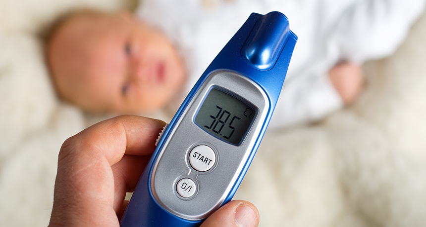
They also said that although in most cases people heal on their own, newborns, children, pregnant women and people with immunodeficiency may have a higher risk of developing severe or even fatal conditions. 

Due to the mild symptoms caused by the monkeypox virus and the lack of large-scale global outbreaks, research into monkeypox vaccines has been relatively limited worldwide, the experts explained.

Meanwhile, as monkeypox and smallpox viruses have extensive serological cross-reactivity, the existing vaccines used for monkeypox prevention are all smallpox vaccines. 

Retrospective studies conducted by the World Health Organization have shown that smallpox vaccine administration has an efficacy of 85 percent in preventing monkeypox. Currently, there are three smallpox vaccines approved for monkeypox prevention in Europe, the US, and Japan, Su Jinfeng, a senior biomedical engineer, told the Global Times. 

Su called for accelerated development of new vaccines to protect those at greater risk and to prevent potential outbreaks. However, the expert admitted that the development of vaccines faces several challenges due to the limited number of monkeypox cases in the country and the dispersed population, which makes it difficult to conduct large-scale clinical trials to assess a vaccine's efficacy. 

"Currently, the US, Japan and European countries have considered this type of vaccine as a reserve drug. China should also accelerate the development of a new smallpox/monkeypox vaccine, not only to prevent the spread of monkeypox outbreaks but also to protect national security and public health from threats of smallpox virus being used as a bioweapon," a vaccine expert who preferred not to be named told the Global Times. 

Given the large genome and complex structure of the monkeypox virus, as well as limited understanding of protective antigens, the development of a protective antigen-based vaccine is challenging, the expert said. Therefore, a better strategy would be to use attenuated live vaccine technology, building upon the existing smallpox vaccine, to develop a safer vaccine in human cells.

As of April 2023, preclinical research on monkeypox vaccines has been conducted primarily by the US and China, said Su. Previous reports have indicated that a total of 14 clinical studies on monkeypox vaccines have been conducted globally.

Currently, three types of vaccine have been approved for the prevention of monkeypox, from Denmark, the US, and Japan. 

Research institutions in China have already started developing monkeypox vaccines, mainly focusing on replication-defective monkeypox attenuated live vaccines and monkeypox mRNA vaccines.

In July, the replication-deficient monkeypox vaccine developed by China National Pharmaceutical Group Corporation (Sinopharm) has passed the clinical trial application with the National Medical Products Administration, making it the earliest domestically developed monkeypox vaccine to enter the clinical research stage in China.

The Chinese mainland has reported 491 new monkeypox cases across 23 provincial-level regions, the country's Center for Disease Control and Prevention confirmed on Wednesday, increasing over fourfold compared to last month. 

According to epidemiological reviews, all cases are male with 96.3 percent of them identified as men who had sex with other men, and the risk of transmission through other contact methods is low.

The majority of cases exhibited typical clinical symptoms including fever, rash, and swollen lymph nodes, with no severe or fatal cases.

Ancient DNA reveals who is in Spain’s ‘pit of bones’ cave

Neandertals hung out in what’s now northern Spain around 430,000 years ago, an analysis of ancient DNA suggests. That’s an earlier Neandertal presence in Europe, by at least 30,000 years, than many researchers had assumed.

Fragments of nuclear DNA from a tooth and partial leg bone discovered at Sima de los Huesos, a chamber deep inside a Spanish cave, resemble corresponding parts of a previously reassembled Neandertal genome, researchers say in a study published online March 14 in Nature.
Not much nuclear DNA survives in such ancient fossils, say paleogeneticist Matthias Meyer of the Max Planck Institute for Evolutionary Anthropology in Leipzig, Germany, and his colleagues. Meyer’s group recovered DNA fragments covering a fraction of 1 percent of the newly recovered Neandertal tooth and leg genomes. Just enough DNA remained to enable comparisons with DNA of a Neandertal woman (SN: 1/25/14, p. 17) and a Denisovan woman (SN: 9/22/12, p. 5). Denisovans are considered close genetic cousins of Neandertals.

The early age for the new genetic finds challenges the idea that fossils from Sima de los Huesos, or pit of bones, come from a species called Homo heidelbergensis. Some researchers have suspected that by around 400,000 years ago, H. heidelbergensis gave rise to evolutionary precursors of both Neandertals and Homo sapiens.
An ancient genetic puzzle has also emerged at Sima de los Huesos. On one hand, nuclear DNA — which passes from both parents to their children — pegs the Spanish hominids as Neandertals. But mitochondrial DNA — typically inherited only from the mother — already extracted from one Sima de los Huesos fossil (SN: 12/28/13, p. 8) and described for a second fossil in the new study has more in common with Denisovans.

Denisovans lived in East Asia at least 44,000 years ago, but their evolutionary history is unknown.

If early Neandertals lived in northern Spain roughly 430,000 years ago, “we have to go back further in time to reach the common ancestor of Neandertals and Denisovans,” Meyer says.
The new genetic data from Sima de los Huesos now suggest that Denisovans split from Neandertals perhaps 450,000 years ago, says paleoanthropologist Chris Stringer of the Natural History Museum in London. Genetic and fossil evidence point to Neandertals and H. sapiens diverging from a common ancestor around 650,000 years ago, he proposes.

But it’s hard to say whether that common ancestor was H. heidelbergensis, Stringer adds. “Research must refocus on fossils from 400,000 to 800,000 years ago to determine which ones might lie on ancestral lineages of Neandertals, Denisovans and modern humans.”

Hominids throughout Eurasia during that time may have shared a mitochondrial DNA pattern observed in Sima de los Huesos Neandertals and Asian Denisovans, Meyer suggests. If that was the case, Neandertals acquired a new form of mitochondrial DNA by interbreeding with modern humans or their direct ancestors from Africa sometime between 430,000 and 100,000 years ago (SN: 3/19/16, p. 6).

Another possibility is that Neandertals traveled to Europe from Asia more than 430,000 years ago, carrying Denisovan mitochondrial DNA with them, says paleogeneticist Carles Lalueza-Fox of the Institute of Evolutionary Biology in Barcelona. Or hybrid descendants of early Neandertals and early Denisovans may have lived at Sima de los Huesos, carrying Denisovan mitochondrial DNA, he speculates.

“We really need more genetic data from Sima de los Huesos, and other sites of that age, to narrow down these scenarios,” Meyer says.

Heat may outpace corals’ ability to cope

Corals are in hot water — and may soon lose their ability to handle the heat.

In Australia’s Great Barrier Reef, most past bouts of warming allowed many corals to adjust their physiology and avoid serious damage. But as waters warm even more, corals could run out of wiggle room, researchers report in the April 15 Science.

“One of the things that we have been striving for is trying to figure out the rate and limit of … physiological adjustments that corals have, how far you can push them,” says marine biologist Stephen Palumbi of Stanford University, who was not involved with the study. Corals may not be able to cope with much more ocean warming, Palumbi says. “I would take this paper as being the first real indication that we have half a degree at most.”
If water temperatures surge quickly, corals may bleach, losing the bacterial residents that provide them with nutrients and oxygen (and color). But if waters warm slightly — less than the roughly 2 degrees Celsius above average heat spike where bleaching begins — and then cool for a brief time before heating up to a greater extent, corals are better prepared to survive the heat. In the lab, corals exposed to this two-step heating process experienced less bleaching and less cell death than corals suffering a high initial heat wave, the researchers found.

“We liken it to the idea of training for a marathon,” says study coauthor Scott Heron, a physical oceanographer with the National Oceanic and Atmospheric Administration’s Coral Reef Watch in College Park, Md. “If they have a little bit of exposure, and then the recovery period after that … they’re better prepared for the race when it comes.”

From 1985 to 2011, around 75 percent of warming events on Great Barrier Reef sites occurred in this stepwise fashion, probably allowing corals to steel themselves and survive warmer waters. But with climate models predicting a 2-degree increase in sea temperatures by the end of the century, warming events could soon push corals past their bleaching point with no chance to prepare.

Computer simulations predicted that as waters grow warmer, reef heat waves will increase overall. But the fraction of such events that could condition corals to withstand bleaching will fall from 75 percent to 22 percent, the team reports. Most reefs that have experienced preconditioning in the past will start losing the ability to prepare when water temperatures increase by 0.5 degrees, the team predicts. Warming trends suggest that the added half degree should appear within 40 years. “If that protective mechanism does get lost going into the future, then what we’ve seen so far as being bad impacts could become worse,” Heron says.

For now, preparation may help some corals survive in warming seas, but reduced carbon emissions will also be required to sustain coral cover throughout the century, the team’s data suggest. Palumbi says these predictions are very important. “If we get a handle on emissions, there are substantial predicted differences in the way that coral populations live in the future,” he says. “We are still in a position to choose how the future of coral reefs works out.”

Cause of mass starfish die-offs is still a mystery

In the summer of 2013, an epidemic began sweeping through the intertidal zone off the west coast of North America. The victims were several species of sea star, including Pisaster ochraceus, a species that comes in orange and purple variants. (It’s also notable because it’s the starfish that provided ecology with the fundamental concept of a keystone species.) Affected individuals appeared to “melt,” losing grip with the rocks to which they were attached — and then losing their arms. This sea star wasting disease, as it is known, soon killed sea stars from Baja California to Alaska.

This wasn’t the first outbreak of sea star wasting disease. A 1978 outbreak in the Gulf of California, for instance, killed so many Heliaster kubinjiisun stars that the once ubiquitous species is now incredibly rare.

These past incidents, though, happened fast and within smaller regions, so scientists had struggled to figure out what had happened. With the latest outbreak happening over such a large — and well-studied — region and period of time, marine biologists have been able to gather more data on the disease than ever before. And they’re getting closer to figuring out just what happened in this latest incident.

One likely factor is the sea star-associated densovirus, which, in 2014, scientists reported finding in greater abundance in starfish with sea star wasting disease than in healthy sea stars. But the virus can’t be the only cause of the disease; it’s found in both healthy and sick sea stars, and it has been around since at least 1942, the earliest year it has been found in museum specimens. So there must be some other factor at play.
Earlier this year, scientists studying the outbreak in Washington state reported in the Proceedings of the Royal Society B thatwarm waters may increase disease progression and rates of death. Studies of California starfish came to a similar conclusion. But a new study, appearing May 4 in PLOS One , finds that may not be true for sea stars in Oregon. Bruce Menge and colleagues at Oregon State University took advantage of their long-term study of Oregon starfish to evaluate what happened to sea stars during the recent epidemic and found that wasting disease increased with cooler , not warmer, temperatures. “Given conflicting results on the role of temperature as a trigger of [sea star wasting disease], it seems most likely that multiple factors interacted in complex ways to cause the outbreak,” they conclude.
What those factors are, though, is still a mystery.

Also unclear is what long-term effects this outbreak will have on Pacific intertidal communities.

In the 1960s, Robert Paine of the University of Washington performed what is now considered a classic experiment. For years, he removed starfish from one area of rock in Makah Bay at the northwestern tip of Washington and left another bit of rock alone as a control. Without the starfish to prey on them, mussels were able to take over. The sea stars, Paine concluded, were a “keystone species” that kept the local food web in control.

If sea star wasting disease has similar effects on the Pacific intertidal food web, Menge and his colleagues write, “it would result in losses or large reductions of many species of macrophytes, anemones, limpets, chitons, sea urchins and other organisms from the low intertidal zone.”

What happens, the group says, may depend on how quickly the disease disappears from the region and how many young sea stars can grow up and start munching on mussels.

Stephen Hawking finds the inner genius in ordinary people

It’s hard to believe that it took reality television this long to get around to dealing with space, time and our place in the cosmos.

In PBS’ Genius by Stephen Hawking, the physicist sets out to prove that anyone can tackle humankind’s big questions for themselves. Each of the series’ six installments focuses on a different problem, such as the possibility of time travel or the likelihood that there is life elsewhere in the universe. With Hawking as a guide, three ordinary folks must solve a series of puzzles that guide them toward enlightenment about that episode’s theme. Rather than line up scientists to talk at viewers, the show invites us to follow each episode’s trio on a journey of discovery.
By putting the focus on nonexperts, Genius emphasizes that science is not a tome of facts handed down from above but a process driven by curiosity. After working through a demonstration of how time slows down near a black hole, one participant reflects: “It’s amazing to see it play out like this.”
The show is a fun approach to big ideas in science and philosophy, and the enthusiasm of the guests is infectious. Without knowing what was edited out, though, it’s difficult to say whether the show proves Hawking’s belief that anyone can tackle these heady questions. Each situation is carefully designed to lead the participants to specific conclusions, and there seems to be some off-camera prompting.

But the bigger message is a noble one: A simple and often surprising chain of reasoning can lead to powerful insights about the universe, and reading about the cosmos pales next to interacting with stand-ins for its grandeur. It’s one thing, for example, to hear that there are roughly 300 billion stars in the Milky Way. But to stand next to a mountain of sand where each grain represents one of those stars is quite another. “I never would have got it until I saw it,” says one of the guests, gesturing to the galaxy of sand grains. “This I get.”

Snot could be crucial to dolphin echolocation

In hunting down delicious fish, Flipper may have a secret weapon: snot.

Dolphins emit a series of quick, high-frequency sounds — probably by forcing air over tissues in the nasal passage — to find and track potential prey. “It’s kind of like making a raspberry,” says Aaron Thode of the Scripps Institution of Oceanography in San Diego. Thode and colleagues tweaked a human speech modeling technique to reproduce dolphin sounds and discern the intricacies of their unique style of sound production. He presented the results on May 24 in Salt Lake City at the annual meeting of the Acoustical Society of America.

Dolphin chirps have two parts: a thump and a ring. Their model worked on the assumption that lumps of tissue bumping together produce the thump, and those tissues pulling apart produce the ring. But to match the high frequencies of live bottlenose dolphins, the researchers had to make the surfaces of those tissues sticky. That suggests that mucus lining the nasal passage tissue is crucial to dolphin sonar.

The vocal model also successfully mimicked whistling noises used to communicate with other dolphins and faulty clicks that probably result from inadequate snot. Such techniques could be adapted to study sound production or echolocation in sperm whales and other dolphin relatives.
Researchers modified a human speech model developed in the 1970s to study dolphin echolocation. The animation above mimics the vibration of lumps of tissue (green) in the dolphin’s nasal passage (black) that are drenched in mucus. Snot-covered tissues (blue) stick together (red) and pull apart to create the click sound.

Jupiter’s stormy weather no tempest in teapot

Jupiter’s turbulence is not just skin deep. The giant planet’s visible storms and blemishes have roots far below the clouds, researchers report in the June 3 Science. The new observations offer a preview of what NASA’s Juno spacecraft will see when it sidles up to Jupiter later this year.

A chain of rising plumes, each reaching nearly 100 kilometers into Jupiter, dredges up ammonia to form ice clouds. Between the plumes, dry air sinks back into the Jovian depths. And the famous Great Red Spot, a storm more than twice as wide as Earth that has churned for several hundred years, extends at least dozens of kilometers below the clouds as well.

Jupiter’s dynamic atmosphere provides a possible window into how the planet works inside. “One of the big questions is what is driving that change,” says Leigh Fletcher, a planetary scientist at the University of Leicester in England. “Why does it change so rapidly, and what are the environmental and climate-related factors that result from those changes?”

To address some of those questions, Imke de Pater, a planetary scientist at the University of California, Berkeley, and colleagues observed Jupiter with the Very Large Array radio observatory in New Mexico. Jupiter emits radio waves generated by heat left over from its formation about 4.6 billion years ago. Ammonia gas within Jupiter’s atmosphere intercepts certain radio frequencies. By mapping how and where those frequencies are absorbed, the researchers created a three-dimensional map of the ammonia that lurks beneath Jupiter’s clouds. Those plumes and downdrafts appear to be powered by a narrow wave of gas that wraps around much of the planet.

The depths of Jupiter’s atmospheric choppiness isn’t too surprising, says Scott Bolton, a planetary scientist at the Southwest Research Institute in San Antonio. “Almost everyone I know would have guessed that,” he says. But the observations do provide a teaser for what to expect from the Juno mission, led by Bolton. The spacecraft arrives at Jupiter on July 4 to begin a 20-month investigation of what’s going on beneath Jupiter’s clouds using tools similar to those used in this study.

The new observations confirm that Juno should work as planned, Bolton says.

By getting close to the planet — just 5,000 kilometers from the cloud tops — Juno will break through the fog of radio waves from Jupiter’s radiation belts that obscures observations made from Earth and limits what telescopes like the Very Large Array can see. But the spacecraft will see only a narrow swath of Jupiter’s bulk at a time. “That’s where ground-based work like the research de Pater has been doing is really essential,” Fletcher says. Observations such as these will let Juno scientists know what’s going on throughout the atmosphere so they can better understand what Jupiter is telling them.

Doctors need better ways to figure out fevers in newborns

Two days after my first daughter was born, her pediatrician paid a house call to examine her newest patient. After packing up her gear, she told me something alarming: “For the next few months, a fever is an emergency.” If we measured a rectal temperature at or above 100.4° Fahrenheit, go to the hospital, she said. Call her on the way, but don’t wait.

I, of course, had no idea that a fever constituted an emergency. But our pediatrician explained that a fever in a very young infant can signal a fast-moving and dangerous bacterial infection. These infections are rare (and fortunately becoming even rarer thanks to newly created vaccines). But they’re serious, and newborns are particularly susceptible.

I’ve since heard from friends who have been through this emergency. Their newborns were poked, prodded and monitored by anxious doctors, in the hopes of quickly ruling out a serious bacterial infection. For infants younger than two months, it’s “enormously difficult to tell if an infant is seriously ill and requires antibiotics and/or hospitalization,” says Howard Bauchner, a pediatrician formerly at Boston University School of Medicine and now editor in chief of the Journal of the American Medical Association.

A new research approach, described in two JAMA papers published in August, may ultimately lead to better ways to find the cause of a fever.

These days, for most (but not all) very young infants, their arrival at a hospital will trigger a workup that includes a urine culture and a blood draw. Often doctors will perform a lumbar puncture, more commonly known as a spinal tap, to draw a sample of cerebrospinal fluid from the area around the spinal cord.

Doctors collect these fluids to look for bacteria. Blood, urine and cerebrospinal fluid are smeared onto culture dishes, and doctors wait and see if any bacteria grow. In the meantime, the feverish infant may be started on antibiotics, just in case. But this approach has its limitations. Bacterial cultivation can take several days. The antibiotics may not be necessary. And needless to say, it’s not easy to get those fluids, particularly from a newborn.

Some scientists believe that instead of looking for bacteria or viruses directly, we ought to be looking at how our body responds to them. Unfortunately, the symptoms of a bacterial and viral infection are frustratingly similar. “You get a fever. You feel sick,” says computational immunologist Purvesh Khatri of Stanford University. Sadly, there are no obvious telltale symptoms of one or the other, not even green snot. In very young infants, a fever might be the only sign that something is amiss.
But more subtle clues could betray the cause of the fever. When confronted with an infection, our immune systems ramp up in specific ways. Depending on whether we are fighting a viral or bacterial foe, different genes turn up their activity. “The immune system knows what’s going on,” Khatri says. That means that if we could identify the genes that reliably get ramped up by viruses and those that get ramped up by bacteria, then we could categorize the infection based on our genetic response.

That’s the approach used by two groups of researchers, whose study results both appear in the August 23/30 JAMA. One group found that in children younger than 2, two specific genes could help make the call on infection type. Using blood samples, the scientists found that one of the genes ramped up its activity in response to a viral infection, and the other responded to a bacterial infection.

The other study looked at immune responses in even younger children. In infants younger than 60 days, the activity of 66 genes measured in blood samples did a pretty good job of distinguishing between bacterial and viral infections. “These are really exciting preliminary results,” says Khatri, who has used a similar method for adults. “We need to do more work.”

Bauchner points out that in order to be useful, “the test would have to be very, very accurate in very young infants.” There’s very little room for error. “Only time will tell how good these tests will be,” he says. In an editorial that accompanied the two studies, he evoked the promise of these methods. If other experiments replicate and refine the results of these studies, he could envision a day in which the parents of a feverish newborn could do a test at home, call their doctor and together decide if the child needs more care.

That kind of test isn’t here yet, but scientists are working on it. The technology couldn’t come soon enough for doctors and parents desperate to figure out a fever.

Endurance training leaves no memory in muscles

Use it or lose it, triathletes.

Muscles don’t have long-term memory for exercises like running, biking and swimming, a new study suggests. The old adage that once you’ve been in shape, it’s easier to get fit again could be a myth, at least for endurance athletes, researchers in Sweden report September 22 in PLOS Genetics.

“We really challenged the statement that your muscles can remember previous training,” says Maléne Lindholm of the Karolinska Institute in Stockholm. But even if muscles forget endurance exercise, the researchers say, other parts of the body may remember, and that could make retraining easier for people who’ve been in shape before.
Endurance training is amazingly good for the body. Weak muscle contractions, sustained over a long period of time — as in during a bike ride — change proteins, mainly ones involved in metabolism. This translates into more energy-efficient muscle that can help stave off illnesses like diabetes, cardiovascular disease and some cancers. The question is, how long do those improvements last?

Previous work in mice has shown that muscles “remember” strength training (SN: 9/11/10, p. 15). But rather than making muscles more efficient, strength-training moves like squats and push-ups make muscles bigger and stronger. The muscles bulk up as they develop more nuclei. More nuclei lead to more production of proteins that build muscle fibers. Cells keep their extra nuclei even after regular exercise stops, to make protein easily once strength training restarts, says physiologist Kristian Gundersen at the University of Oslo in Norway. Since endurance training has a different effect on muscles, scientists weren’t sure if the cells would remember it or not.
To answer that question, Lindholm’s team ran volunteers through a 15-month endurance training experiment. In the first three months, 23 volunteers trained four times a week, kicking one leg 60 times per minute for 45 minutes. Volunteers rested their other leg. Lindholm’s team took muscle biopsies before and after the three-month period to see how gene activity changed with training. Specifically, the scientists looked for changes in the number of mRNAs (the blueprints for proteins) that each gene was making. Genes associated with energy production showed the greatest degree of change in activity with training.
At a follow-up, after participants had stopped training for nine months, scientists again biopsied muscle from the thighs of 12 volunteers, but didn’t find any major differences in patterns of gene activity between the previously trained legs and the untrained legs. “The training effects were presumed to have been lost,” says Lindholm. After another three-month bout of training, this time in both legs, the researchers saw no differences between the previously trained and untrained legs.
While this study didn’t find muscle memory for endurance — most existing evidence is anecdotal — it still might be easier for former athletes to get triathalon-ready, researchers say. The new result has “no bearing on the possible memory in other organ systems,” Gundersen says. The heart and cardiovascular system could remember and more easily regain previous fitness levels, for example, he says.

Even within muscle tissue, immune cells or stem cells could also have some memory not found in this study, says molecular exercise physiologist Monica Hubal of George Washington University in Washington, D.C. Lindholm adds that well-trained connections between nerves and muscles could also help lapsed athletes get in shape faster than people who have never exercised before. “They know how to exercise, how it’s supposed to feel,” Lindholm says. “Your brain knows exactly how to activate your muscles, you don’t forget how to do that.”

Primitive signs of emotions spotted in sugar-buzzed bumblebees

To human observers, bumblebees sipping nectar from flowers appear cheerful. It turns out that the insects may actually enjoy their work. A new study suggests that bees experience a “happy” buzz after receiving a sugary snack, although it’s probably not the same joy that humans experience chomping on a candy bar.

Scientists can’t ask bees or other animals how they feel. Instead, researchers must look for signs of positive or negative emotions in an animal’s decision making or behavior, says Clint Perry, a neuroethologist at Queen Mary University of London. In one such study, for example, scientists shook bees vigorously in a machine for 60 seconds — hard enough to annoy, but not hard enough to cause injury — and found that stressed bees made more pessimistic decisions while foraging for food.
The new study, published in the Sept. 30 Science, is the first to look for signs of positive bias in bee decision making, Perry says. His team trained 24 bees to navigate a small arena connected to a plastic tunnel. When the tunnel was marked with a blue “flower” (a placard), the bees learned that a tasty vial of sugar water awaited them at its end. When a green “flower” was present, there was no reward. Once the bees learned the difference, the scientists threw the bees a curveball: Rather than being blue or green, the “flower” had a confusing blue-green hue.

Faced with the ambiguous color, the bees appeared to dither, meandering around for roughly 100 seconds before deciding whether to enter the tunnel. Some didn’t enter at all. But when the scientists gave half the bees a treat — a drop of concentrated sugar water — that group spent just 50 seconds circling the entrance before deciding to check it out. Overall, the two groups flew roughly the same distances at the same speeds, suggesting that the group that had gotten a treat first had not simply experienced a boost in energy from the sugar, but were in a more positive, optimistic state, Perry says.

In a separate experiment, Perry and colleagues simulated a spider attack on the bees by engineering a tiny arm that darted out and immobilized them with a sponge. Sugar-free bees took about 50 seconds longer than treated bees to resume foraging after the harrowing encounter.

The researchers then applied a solution to the bees’ thoraxes that blocked the action of dopamine, one of several chemicals that transmit rewarding signals in the insect brain. With dopamine blocked, the effects of the sugar treat disappeared, further suggesting that a change in mood, and not just increased energy, was responsible for the bees’ behavior.

The results provide the first evidence for positive, emotion-like states in bees, says Ralph Adolphs, a neuroscientist at Caltech. Yet he suspects that the metabolic effects of sugar did influence the bees’ behavior.
Geraldine Wright, a neuroethologist at Newcastle University in England, shares that concern. “The data reported in the paper doesn’t quite convince me that eating sucrose didn’t change how they behaved, even though they say it didn’t affect flight time or speed of flight,” she says. “I would be very cautious in interpreting the responses of bees in this assay as a positive emotional state.”